
Mouse Secondary Antibodies

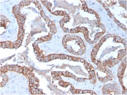
Biotium Cytokeratin 15 (Esophageal Squamous Cell Carcinoma Marker)(KRT15/2957), CF740 conjugate, 0.1mg/mL
Keratin 15 is a type I keratin which is expressed only in basal keratinocytes in stratified epithelia and does not appear to have a natural type II expression partner. Keratin 15 is down regulated in activated keratinocytes. Cytokeratin 15 is a specific marker of stem cells of the hair-follicle bulge and may be a useful marker for diagnosis between basal cell carcinoma (BCC) and trichoepithelioma. Trichoblastoma are benign neoplasms of follicular differentiation frequently found in nevus sebaceous. Many morphologic features are shared with nodular basal cell carcinoma, sometimes rendering a diagnosis difficult. Trichoblastoma and BCC show variable expression of Cytokeratin 15 and Cytokeratin 19, and absence of hair keratins. Primary antibodies are available purified, or with a selection of fluorescent CF Dyes and other labels. CF Dyes offer exceptional brightness and photostability. Note: Conjugates of blue fluorescent dyes like CF405S and CF405M are not recommended for


Biotium Napsin A (Lung Adenocarcinoma Marker)(NAPSA/1238 + NAPSA/1239), CF740 conjugate, 0.1mg/mL
Napsin is a pepsin-like aspartic proteinase connected with maturation of surfactant protein B.There are two closely related napsins, napsin A and napsin B. Napsin A is expressed as a single chain protein. Immunohistochemical studies revealed high expression levels of napsin A in human lung and kidney but low expression in spleen. Napsin A is expressed in type II pneumocytes and in adenocarcinomas of lung. The high specificity expression of napsin A in adenocarcinomas of lung is useful to distinguish primary lung adenocarcinomas from adenocarcinomas of other organs.
Biotium Mucin 3(MUC3/1154), CF568 conjugate, 0.1mg/mL
This antibody recognizes a protein of HMW, identified as mucin 3 glycoprotein (MUC3). This MAb shows no cross-reaction with human milk fat globule membranes, MUC1, or MUC2. The Mucins are a family of highly glycosylated, secreted proteins with a basic structure consisting of a variable number of tandem repeats (VNTRs) encoded by 60 base pairs (Mucin 1), 69 base pairs (Mucin 2) and 51 base pairs (Mucin 3). The number of repeats is highly polymorphic and varies among different alleles. Mucin 1 proteins are expressed as type I membrane proteins in addition to secreted forms. Mucin 1 is aberrantly expressed in epithelial tumors including breast carcinomas. Mucin 2 coats the epithelia of the intestines and airways and is associated with colonic tumors. Mucin 3 is a major component of various mucus gels and is broadly expressed in normal and tumor cells.Primary antibodies are available purified, or with a selection of fluorescent CF Dyes and other labels. CF Dyes offer exceptional bri
Biotium Galectin-1 / Human Placental Lactogen (hPL) (GAL1/1831), Biotin conjugate, 0.1mg/mL
Galectin-1 is a member of the beta-galactoside-binding family and is a dimeric protein of 14 kD participating in a variety of normal and pathological processes, including cancer progression. Galectin-1 can affect the proliferation of normal and malignant cells. Inhibition of cell growth is observed in a lactose-dependent manner as lower concentrations of the lectin stimulate cell proliferation. Galectin-1 may also be implicated in the induction of apoptosis of activated T cells through the binding of exogenous galectin-1 to CD45 molecules present on the surface of lymphocytes. Galectin-1, reported to be present either at the surface of cancer cells or accumulated around these cells could act as an immunological shield to protect against a T cell immune response and provide an advantage for survival.Primary antibodies are available purified, or with a selection of fluorescent CF Dyes and other labels. CF Dyes offer exceptional brightness and photostability. Note: Conjugates
Biotium TIMP2 (Tissue Inhibitor of Metalloproteinase 2) (3A4), CF568 conjugate, 0.1mg/mL
This antibody recognizes a protein of 21 kDa, identified as tissue inhibitor of metalloproteinases-2 (TIMP-2). It is closely related to TIMP-1 and shows the highest binding affinity to both the latent (pro) and active forms of 72 kDa Type IV collagenase (also known as MMP-2 or gelatinase A). It also has affinity for the active form of 92 kDa Type IV collagenase (also known as MMP-9 or gelatinase B). TIMP-2 inhibits the proteolytic invasiveness of tumor cells and normal placental trophoblast cells.Primary antibodies are available purified, or with a selection of fluorescent CF Dyes and other labels. CF Dyes offer exceptional brightness and photostability. Note: Conjugates of blue fluorescent dyes like CF405S and CF405M are not recommended for detecting low abundance targets, because blue dyes have lower fluorescence and can give higher non-specific background than other dye colors.
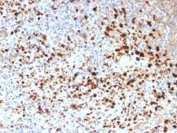
Biotium HSV1 (Herpes Simplex Virus Type I)(HSV1/1934), 1mg/mL
The antibody reacts with HSV type 1 specific antigen. It is suitable for detection of HSV in human cellular material obtained from superficial lesions or biopsies and for the early identification of HSV in infected tissue cultures. The herpes simplex virus (HSV) (also known as cold sore, night fever or fever blister) is a virus that causes a contagious disease. There are two main types of Herpes Simplex Virus (HSV), 1 and 2. The HSV-1 strain generally appears in the orafacial organs. HSV2 usually resides in the sacral ganglion at the base of the spine. All herpes viruses are morphologically identical: they have a large double-stranded DNA genome and the virion consists of an icosahedral nucleo-capsid, which is surrounded by a lipid bilayer envelope.Primary antibodies are available purified, or with a selection of fluorescent CF Dyes and other labels. CF Dyes offer exceptional brightness and photostability. Note: Conjugates of blue fluorescent dyes like CF405S and CF405M
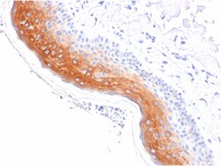
Biotium Cytokeratin 1 (Suprabasal Keratinocyte Marker)(KRT1/1840), 1mg/mL
Keratins are a family of intermediate filament proteins that assemble into filaments through forming heterodimers of one type I keratin (keratins 9 to 23) and one type II keratin (keratins 1 to 8). Keratins demonstrate tissue and differentiation specific expression profiles. Cytokeratin 1 is a hetero-tetramer of two type I as well as two type II keratins, keratin-10 (KRT10). Keratin 1 is differentiation specific keratin that is one of the predominant keratins in suprabasal keratinocytes in stratified epithelia. Mutations in keratin 1 cause epidermolytic hyperkeratosis. Primary antibodies are available purified, or with a selection of fluorescent CF Dyes and other labels. CF Dyes offer exceptional brightness and photostability. Note: Conjugates of blue fluorescent dyes like CF405S and CF405M are not recommended for detecting low abundance targets, because blue dyes have lower fluorescence and can give higher non-specific background than other dye colors.
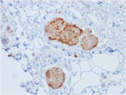
Biotium Chromogranin A / CHGA (Neuroendocrine Marker)(rCHGA/777), CF568 conjugate, 0.1mg/mL
Chromogranin A is present in neuroendocrine cells throughout the body, including the neuroendocrine cells of the large and small intestine, adrenal medulla and pancreatic islets. It is an excellent marker for carcinoid tumors, pheochromocytomas, paragangliomas, and other neuroendocrine tumors. Co-expression of chromogranin A and neuron specific enolase (NSE) is common in neuroendocrine neoplasms. Reportedly, co-expression of certain keratins and chromogranin indicates neuroendocrine lineage. The presence of strong anti-chromogranin staining and absence of anti-keratin staining should raise the possibility of paraganglioma. The co-expression of chromogranin and NSE is typical of neuroendocrine neoplasms. Most pituitary adenomas and prolactinomas readily express chromogranin. Primary antibodies are available purified, or with a selection of fluorescent CF Dyes and other labels. CF Dyes offer exceptional brightness and photostability. Note: Conjugates of blue fluorescent dyes
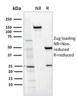
Biotium CD36 (Platelet & Microvessel Marker)(GPIIIb/1654), CF647 conjugate, 0.1mg/mL
Recognizes a protein of 80 kDa-90 kDa, identified as CD36. It is expressed on platelets, monocytes and macrophages, microvascular endothelial cells, erythrocyte precursors, mammary epithelial cells, and some macrophage derived dendritic cells. CD36 acts as a receptor for thrombospondin (TSP), collagen types I, IV and V, P. falciparum malaria-infected erythrocytes, and sickle erythrocytes. It also functions as a scavenger receptor, mediating macrophage uptake of oxidized low-density lipoprotein (LDL) and recognition of apoptotic polymorphonuclear leukocytes (PMN). CD36 plays a role in platelet aggregation, macrophage foam cell development, inflammation, and the tissue ischemia observed in sickle cell disease and cerebral malaria. Note that 1-4% of Japanese and East Asia population lack CD36. Primary antibodies are available purified, or with a selection of fluorescent CF Dyes and other labels. CF Dyes offer exceptional brightness and photostability. Note: Conjugates of blue
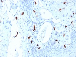
Biotium Tryptase (Mast Cell Marker) (TPSAB1/1961), CF740 conjugate, 0.1mg/mL
Tryptases comprise a family of trypsin-like serine proteases (peptidase family S1). Tryptases are stored in mast cell secretory granules and basophils. Mast cells are connective tissue cells derived from blood-forming tissues that line arterial walls and secrete substances, which mediate inflammatory and immune responses. Tryptases are released into the extracellular environment and are resistant to all known endogenous proteinase inhibitors. This antibody reacts with mast cells distributed in skin, synovium, lung, and heart. This antibody does not bind with any other cell type. Human mast cell tryptase is considered to be an important marker of mast cell activation and is an important mediator of inflammation. Mastocytosis is a term collectively used for a group of disorders in which there is abnormal accumulation of mast cells in one or multiple organs. Anti-tryptase, combined with anti-CD2, anti-CD25, and anti-CD117, can be useful in identifying reactive mast cell hyperpla
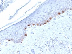
Biotium Tyrosinase-Related Protein-1 (TYRP-1) (Melanoma Marker) (TYRP1/1986), CF488A conjugate, 0.1mg/mL
This antibody reacts with a 75 kDa melanocyte-specific gene product, identified as Tyrosinase-related protein-1 (TRP-1). It is involved in melanin synthesis. TRP1 is present on the melanosomal membranes of melanoma, normal melanocytes and nevi. Recent evidence suggests that TRP-1 is involved in maintaining stability of tyrosinase protein and modulating its catalytic activity. TRP-1 is also involved in maintenance of melanosome ultrastructure and affects melanocyte proliferation and cell death.Primary antibodies are available purified, or with a selection of fluorescent CF Dyes and other labels. CF Dyes offer exceptional brightness and photostability. Note: Conjugates of blue fluorescent dyes like CF405S and CF405M are not recommended for detecting low abundance targets, because blue dyes have lower fluorescence and can give higher non-specific background than other dye colors.
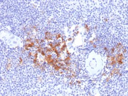
Biotium TRAF1 (TNFR-Associated Factor 1) (Lymphomatoid Papulosis Marker)(TRAF1/2770), CF594 conjugate, 0.1mg/mL
This MAb recognizes a protein of 52 kDa, which is identified as TNFR1 (TNFR-associated factor 1). CD30-positive lymphoproliferations of the skin comprise 30% of all primary cutaneous T-cell lymphomas (CTCLs). Besides borderline cases this group includes lymphomatoid papulosis (LyP) and primary cutaneous anaplastic large T-cell lymphoma (cALCL). Although the two entities overlap clinically, histopathologically, immunopathologically and genetically, they differ considerably in their prognosis. In particular, common feature of both cases is histologically the presence of atypical lymphoid CD30-positive T blasts and genetically a clonal T-cell-receptor rearrangement. However, both cases differ considerably in their clinical course: Lesions of LyP regress spontaneously, whereas those of cALCL persist and may progress and spread. Moreover, LyP patients do not benefit from an aggressive radio- and/or chemotherapeutic approach, in contrast to patients with cALCL. Besides, LyP and cAL
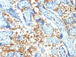
Biotium Spectrin Alpha 1 (Erythrocyte Marker) (SPTA1/1810), 0.2mg/mL
Spectrin is an actin crosslinking and molecular scaffold protein that links the plasma membrane to the actin cytoskeleton, and functions in the determination of cell shape, arrangement of transmembrane proteins, and organization of organelles. Spectrins function as membrane organizers and stabilizers, composed of non-homologous α andβ chains, which aggregate side-to-side in an anti-parallel fashion to form dimers, tetramers and higher polymers. Spectrin α I and spectrinβ I are present in erythrocytes, whereas spectrin α II (also designated fodrin α) and spectrinβ II (also designated fodrinβ) are present in other somatic cells. The spectrin tetramers in erythrocytes act as barriers to lateral diffusion, but spectrin dimers seem to lack this function.Primary antibodies are available purified, or with a selection of fluorescent CF Dyes and other labels. CF Dyes offer exceptional brightness and photostability. Note: Conjugates of blue fluorescent dyes like CF405S
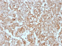
Biotium DOG-1 / TMEM16A (Gastrointestinal Stromal Tumor Marker)(rDG1/447), CF568 conjugate, 0.1mg/mL
Expression of DOG-1 protein is elevated in the gastrointestinal stromal tumors (GIST's), c-kit signaling-driven mesenchymal tumors of the GI tract. DOG-1 is rarely expressed in other soft tissue tumors, which, due to appearance, may be difficult to diagnose. Immunoreactivity for DOG-1 has been reported in 97. 8 percent of scorable GIST's, including all c-kit negative GIST's. Overexpression of DOG-1 has been suggested to aid in the identification of GISTs, including Platelet-Derived Growth Factor Receptor Alpha mutants that fail to express c-kit antigen. The overall sensitivity of DOG1 and c-kit in GIST's is nearly identical: 94. 4% vs. 94. 7%. Primary antibodies are available purified, or with a selection of fluorescent CF Dyes and other labels. CF Dyes offer exceptional brightness and photostability. Note: Conjugates of blue fluorescent dyes like CF405S and CF405M are not recommended for detecting low abundance targets, because blue dyes have lower fluorescence and can
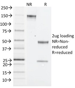
Biotium NK1.1 / CD161c / Klrb1c, Mouse(PK136), CF568 conjugate, 0.1mg/mL
NK1. 1 is a type II integral membrane glycoprotein with a C-type lectin domain and is encoded by theKlrb1c/NKR-P1Cgene. It is predominantly expressed as a disulfide-linked homodimer on NK cells however; it is also expressed on NK-T cells, a rare population of T lymphocytes. NK1. 1 is expressed in some mouse strains, including C57BL/6, FVB/N, and NZB, but not AKR, BALB/c, CBA/J, C3H, DBA/1, DBA/2, NOD, SJL, and 129. Expression of NKR-P1C antigen has been correlated with lysis of tumor cells in vitro and rejection of bone marrow allograftsin vivo. NK-1. 1 has also been shown to play a role in NK cell activation, IFN- production, and cytotoxic granule release. NK-1. 1 is commonly used as a mouse NK cell marker. Primary antibodies are available purified, or with a selection of fluorescent CF Dyes and other labels. CF Dyes offer exceptional brightness and photostability. Note: Conjugates of blue fluorescent dyes like CF405S and CF405M are not recommended for detecting low abu